Séminaire mercredi 22 mai 2024
 Unraveling the nanoscale organisation of marine calcifiers using advanced microscopy and spectroscopy
Roland Kröger, professeur à l’université d’York
Mercredi 22 mai à 13 heures en salle 303
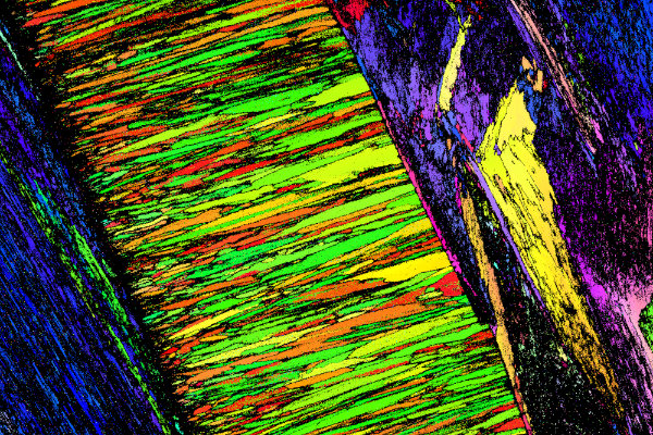
Key to comprehending how marine calcifying invertebrates create and assemble their skeletons and shells is a detailed study of the nanoscale level formation of calcium carbonate based building blocks. Advanced microscopy and spectroscopy techniques such as electron microscopy in conjunction with electron back-scattered diffraction and energy dispersive X-ray spectroscopy as well as Raman spectroscopy allow to obtain detailed insights into both the nanostructure and composition of these structures.
This presentation discusses the application of these methods to a variety of invertebrate calcifiers including corals, coccolithophore algae, abalone sea snails and scallop bivalves. A multimodal approach across the length scales proves the most insightful way to investigate e.g. the 3D arrangement of calcite nanocrystals in the spiral spicules of Clavigera rhabsosphera, the intricate organisation of aragonite platelets in nacre including nanoscale occlusions believed to be associated with mineralisation controlling proteins, the role of nanocrystal nucleation and growth for the skeleton formation in stony corals and the structure of the myostracum in Pecten maximus scallops, which plays a key role in the attachment of the adductor muscle to the bivalve shell. On the basis of these examples a quantitative analysis of the obtained results is undertaken to improve the understanding of the impact of environmental conditions on calcification processes in the marine environment.
- extrait:
- lien_externe:
- kc_data:
- a:8:{i:0;s:0:"";s:4:"mode";s:0:"";s:3:"css";s:0:"";s:9:"max_width";s:0:"";s:7:"classes";s:0:"";s:9:"thumbnail";s:0:"";s:9:"collapsed";s:0:"";s:9:"optimized";s:0:"";}
- kc_raw_content:
- Unraveling the nanoscale organisation of marine calcifiers using advanced microscopy and spectroscopy
Roland Kröger, professeur à l’université d’York
Mercredi 22 mai à 13 heures en salle 303
Key to comprehending how marine calcifying invertebrates create and assemble their skeletons and shells is a detailed study of the nanoscale level formation of calcium carbonate based building blocks. Advanced microscopy and spectroscopy techniques such as electron microscopy in conjunction with electron back-scattered diffraction and energy dispersive X-ray spectroscopy as well as Raman spectroscopy allow to obtain detailed insights into both the nanostructure and composition of these structures.
This presentation discusses the application of these methods to a variety of invertebrate calcifiers including corals, coccolithophore algae, abalone sea snails and scallop bivalves. A multimodal approach across the length scales proves the most insightful way to investigate e.g. the 3D arrangement of calcite nanocrystals in the spiral spicules of Clavigera rhabsosphera, the intricate organisation of aragonite platelets in nacre including nanoscale occlusions believed to be associated with mineralisation controlling proteins, the role of nanocrystal nucleation and growth for the skeleton formation in stony corals and the structure of the myostracum in Pecten maximus scallops, which plays a key role in the attachment of the adductor muscle to the bivalve shell. On the basis of these examples a quantitative analysis of the obtained results is undertaken to improve the understanding of the impact of environmental conditions on calcification processes in the marine environment.
- titre:
- intervenant:
- date: